Researchers Creates New Programmed Shape-Morphing Scaffolds Enabling Facile 3D Endothelialization
Date:30-05-2018 | 【Print】 【close】
A research team led by Dr. DU Xuemin at the Shenzhen Institutes of Advanced Technology (SIAT), Chinese Academy of Sciences has created a new shape-morphing scaffold, enabling programmed deformation from 2D planar cell-laden structure to well-defined 3D tubular shape recently, which facilitated the facile 3D endothelialization of small-diameter vascular grafts. The paper entitled “Programmed Shape-Morphing Scaffolds Enabling Facile 3D Endothelialization” has been published in Advanced Functional Materials.
Cardiovascular disease is now the No. 1 cause of death globally according to the statistic from the World Health Organization, more than 17.5 million patients died from it every year.
Coronary artery bypass grafting (CABG) is one of most effective approaches for treating severe cardiovascular disease.
However, the patients undergoing CABG still confront high risks of transplantation surgery and the potential complications caused by the compliance mismatch.
In recent years, tissue engineering has emerged, holding promise to construct functional vascular analogs for treating cardiovascular disease. Nevertheless, the 3D endothelialization remains greatly challengeable for tissue engineered vascular grafts (TEVGs), particularly small-diameters ones (diameter < 5 mm) suiting for CABG, which is the primary problem of TEVGs upon implantation.
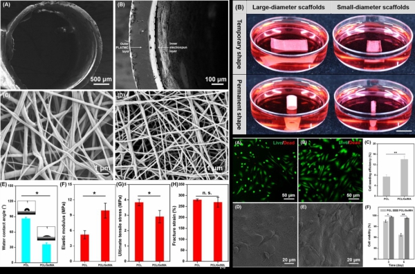
Figure 1. Physical characteristics of shape-morphing scaffolds, and the shape-morphing scaffolds with seeded HUVECs.
For addressing the problem of 3D endothelialization of TEVGs, the research team led by Dr. DU Xuemin designed and developed a novel scaffold, consisting of two layers with combinations of shape memory polymer and electrospun membrane.
By employing the unique property of shape memory polymer, the scaffold could deform from 2D planar shape to well-defined 3D tubular shape at the physiological temperature (37 °C). The endothelial cells seeded onto the electrospun membrane of the planar bilayer scaffold with homogenous and firm cell attachment could be therefore conveniently converted to the vascular-like structure with predetermined tubular shape, where desirable 3D spatial organization of endothelial cells onto the lumen of the scaffold was achieved.
This study found that the 3D cultured endothelial cells on the novel shape-morphing scaffold could form biomimetic cell-scaffold and cell-cell interactions, effectively promoting the formation of confluent endothelial monolayer and 3D endothelialization of TEVGs.
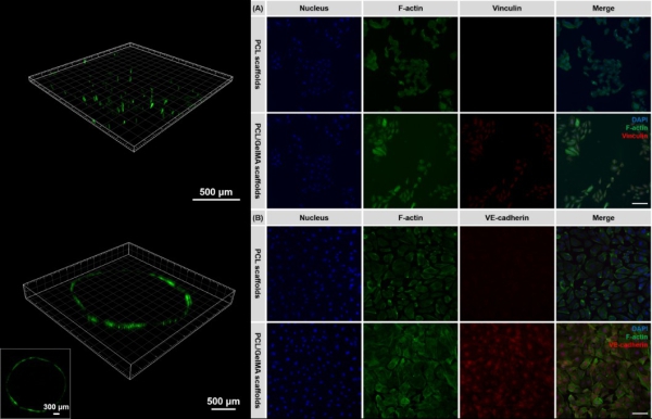
Figure 2. 2D organization of HUVECs (green color, stained by Calcein AM) cultured on the bilayer scaffold, and the immunochemical staining of HUVECs incubated on the different types of shape-morphing scaffolds.
This research offers not only the new route to creat TEVGs enabling facile 3D endothelialization, but also a potential in vitro endothelium model for the screening of drugs for cardiovascular treatment. “Also, we hope that the universal strategy developed in this study by combining smart materials and conventional tissue engineering scaffolds can be extended to engineer complex cell-scaffold constructs mimicking the complicated anatomy of various tissues and organs through the on-demand programmed deformation.” said Dr. DU Xuemin.
CONTACT:
ZHANG Xiaomin
Email: xm.zhang@siat.ac.cn
Tel: 86-755-86585299