New Full Field-of-view Photoacoustic/Ultrasonic Endoscopic System for Early Detection of Gastrointestinal Cancer
Date:13-06-2018 | 【Print】 【close】
Scientists have developed a new endoscopic system elegantly combining photoacoustic and ultrasonic imaging for gastrointestinal (GI) tract, which has an enormous potential of detecting tumors at the early stage and is still a big challenge for conventional endoscopes.
With a full field-of-view imaging capability, the system can provide both high-resolution morphological and high-sensitive functional information. Additionally, the size of system is totally compact to fit in the instrumental channel, which makes it easier to be applied to clinical diagnosis in the near future.
Globally, GI cancer is the third most common types of cancer. 90% of the patients can survive in first five years with proper diagnosis at the early stage of GI cancer.
Many GI diseases, such as tumor and inflammation, are associated with vessel distribution and blood oxygen saturation status. Abnormal vessel distribution and blood oxygen metabolic rate always occurs before the morphological disorder of GI tract at the early stage of a tumor.
Functional photoacoustic endoscope has the potential to measure the blood oxygen metabolic rate, present the vessel morphology and image the 3D vessels distribution in the subcutaneous mucous membrane level, which are associated with GI diseases and always occurs before morphological disorder of GI tract at the early stage of tumor and inflammation.
A research team led by Prof. SONG Liang and Dr. GONG Xiaojing at the Shenzhen Institutes of Advanced Technology (SIAT) of the Chinese Academy of Sciences, developed this photoacoustic/ultrasonic dual-modality endoscopic system and a corresponding miniaturized, encapsulated imaging catheter, the paper “In vivo photoacoustic/ultrasonic dual-modality endoscopy with a miniaturized full field-of-view catheter” was published in Journal of Biophotonics.
The catheter, which is with a 2.5-mm diameter, is compatible with the 2.8-mm instrumental channel of a conventional clinical optical endoscope. The team demonstrated the imaging ability by performing photoacoustic and ultrasonic imaging with a full 360-degree field-of-view. Benefiting from the design of the system and catheter, it is the first time to accomplish in vivo full field-of-view photoacoustic endoscopic imaging in a small animal gastrointestinal tract.
"Using this system, we demonstrate in vivo 3D endoscopic photoacoustic/ultrasonic imaging of the colorectum of a healthy SD rat, by depicting vasculature and morphology of the gastrointestinal tract. The significantly improved imaging field of view, reduced catheter size, high-quality imaging results suggest that the developed dual-modality endoscopy has a great potential to be translated into a broad range of clinical applications in gastroenterology.” Said Prof. SONG Liang.
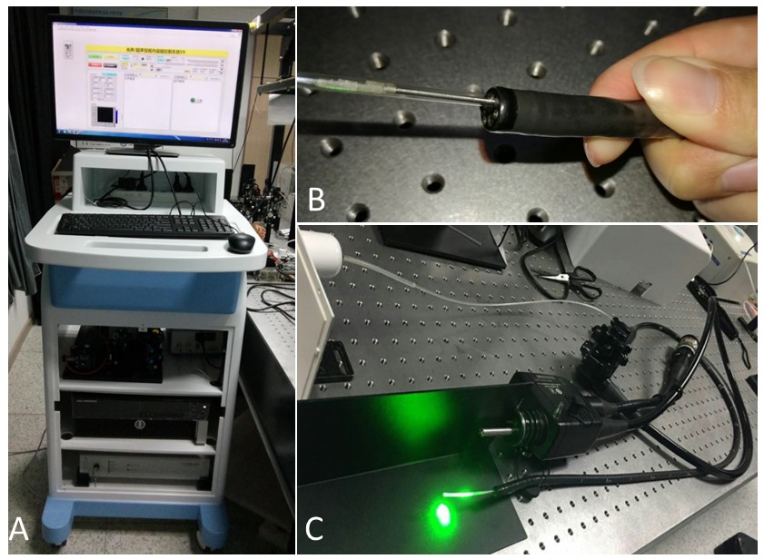
Figure 1. A: Photograph of the endoscopic system. B: The catheter, which is with a 2.5-mm diameter, is compatible with the 2.8-mm instrumental channel of a conventional clinical optical endoscope. C: The 2-m long catheter is working with an endoscope.
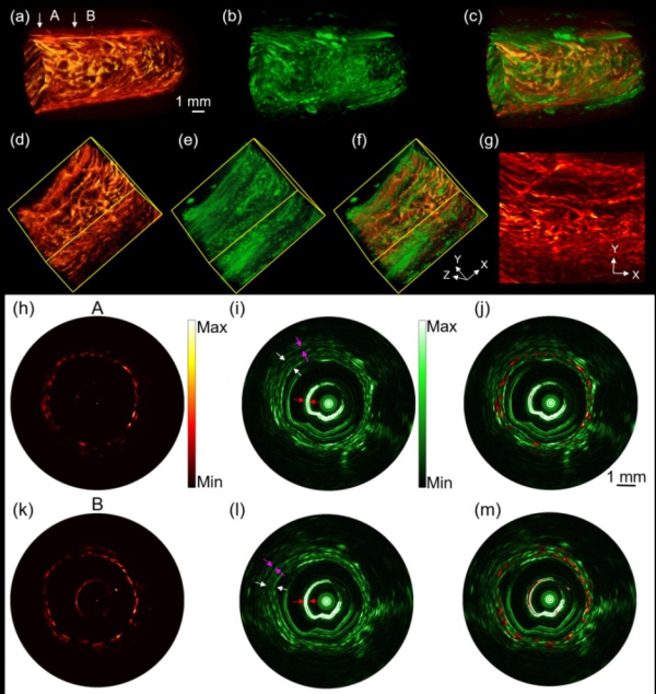
Figure 2. Imaging results of a healthy rat rectum. (A) 3D photoacoustic and (B) ultrasound images of the rectum, (C) Fused photoacoustic and ultrasound images, (D-F) Unfolded images of (A-C),respectively, (G) A maximum amplitude projection (MAP) image of (D), (H-M) Photoacoustic, ultrasound, and fused B-Scan images of “A” and “B” cross-section (labeled by the arrows in A), respectively. Pink arrow: rectal circular muscle; red arrow: protective sheath; white arrow: rectum
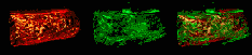
Figure 3. Imaging results of a healthy rat rectum. (A) 3D photoacoustic and (B) ultrasound images of the rectum, (C) Fused photoacoustic and ultrasound images, (D-F) Unfolded images of (A-C),respectively, (G) A maximum amplitude projection (MAP) image of (D), (H-M) Photoacoustic, ultrasound, and fused Bscan images of “a” and “b” cross-section (labeled by the arrows in A), respectively.
Pink arrow: rectal circular muscle; red arrow: protective sheath; white arrow: rectum