Scientists Uncover Key Brain Mechanism in Salience Processing
Date:26-10-2018 | 【Print】 【close】
Scientists have discovered a new brain mechanism underlying salience processing that controls associative learning.
The salience of a stimulus is the state of being noticeable or important. Saliency detection is a key brain mechanism that facilitates learning and survival by enabling organisms to focus their limited attention resources on the most important event. The brain circuits underlying saliency detection had not previously been well understood.
A study carried out by Prof. ZHU Yingjie from the Shenzhen Institutes of Advanced Technology (SIAT) of the Chinese Academy of Sciences, in collaboration with Professor CHEN Xiaoke from Stanford University, uncovered the brain mechanism underlying salience processing. The results were published in Science as a research article on October 26.
By using fiber photometry and single-unit recording, the researchers found that neurons in the periventricular nucleus of the thalamus (PVT) are robustly activated by a variety of salient stimuli, including novel stimuli, reinforcing stimuli and their predicting cues. The responses were proportional to stimulus intensity, and a larger reward (punishment) could evoke a larger PVT response.
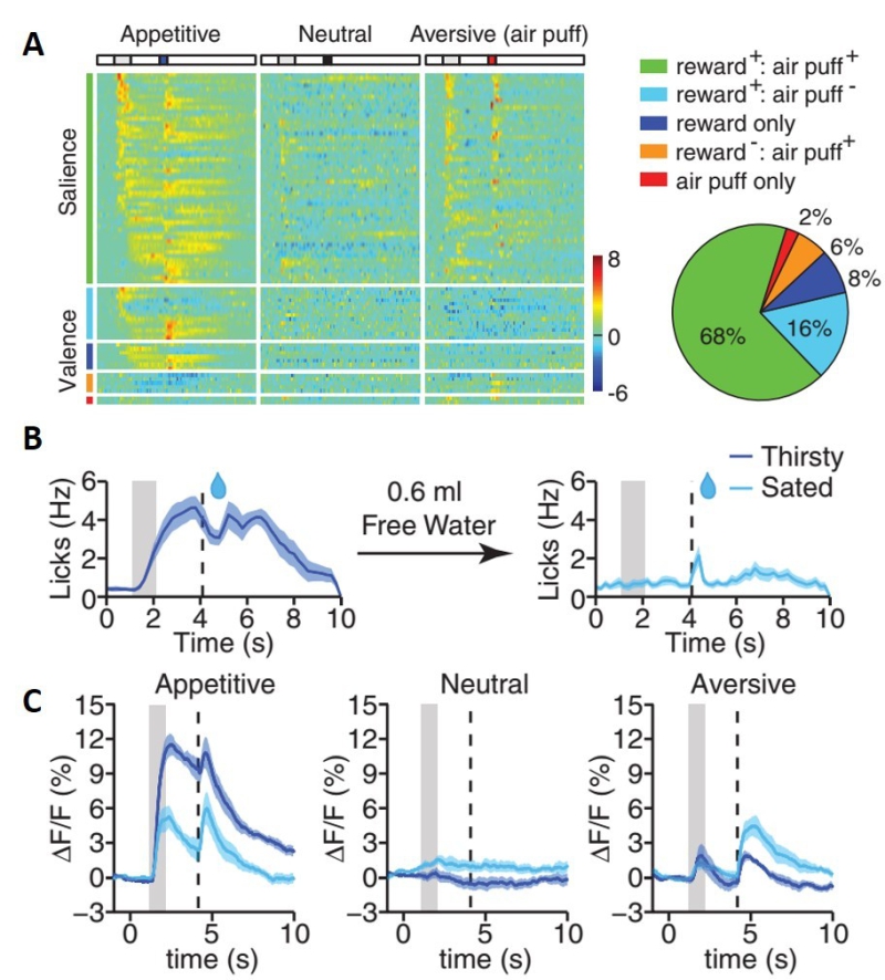
The salience of stimuli is not only determined by physical properties and behavioral relevance of the stimuli, but also depends on the internal homeostatic state of the animal. For example, water is a very salient stimulus when an animal is thirsty, but its saliency should decrease when an animal is in a sated state. Researchers found that PVT neuron responses to water and their predicting cue decrease after an animal had consumed a significant amount of water.

Fig. PVT tracks context-dependent salience (A) Z score heat maps (left) and pie chart (right) for all task responding neurons identified by means of in vivo single-unit recording during Pavlovian tasks. Each row in the heat maps represents responses from the same neuron to different stimuli. (B) Mean lick rate after odor cue in thirsty (left) and sated (right) state. (C) Mean photometric traces of PVT responses in appetitive, neutral, and aversive test in thirsty and sated state.
The salience of stimuli also depends on the external environment of the animal. For example, the saliency of water should decrease when a predator is present. By modulating the behavioral context, researchers showed that the PVT could dynamically track the salience of stimuli upon changes to the internal homeostatic state and external environment.
Researchers found that PVT salience is important for associative learning. Optogenetic suppression of the PVT during learning could significantly disrupt both appetitive and aversive learning.
The PVT can also be activated by internally generated emotional salience such as the disappointment associated with the omission of an expected reward. This omission response is important for extinction learning. Optogenetic suppression of the PVT can slow down extinction learning.
Prof. Robert C. Malenka, deputy director of the Stanford Neurosciences Institute, lauded the research by calling it “a very sophisticated and elegant study of how the brain processes and encodes the motivational importance or ‘salience’ of behaviorally significant stimuli.” He expressed confidence that scientists studying brain mechanisms underlying adaptive and pathological behaviors in humans would “pay close attention to this work and begin exploring the role of this critical brain region, the PVT. “