Researchers Demonstrate the Spatiotemporal Dynamic Changes in Brain Lipids
Date:19-10-2022 | 【Print】 【close】
Ischemic stroke is a leading cause of permanent disability worldwide. Reperfusion therapy (re-opening the artery) with the thrombolytic agent recombinant tissue-type plasminogen activator (rtPA) or mechanical thrombectomy is the most effective treatment for ischemic stroke currently. However, a large proportion of stroke patients remain severely disabled even after receiving timely reperfusion therapy.
It remains unclear how reperfusion therapy results in secondary injury to the brain tissue and whether different reperfusion therapies induce differential effects.
Lipids are essential small molecule compounds for brain function and homeostasis, in which they can not only act as structural components of the cell membrane and fuel for energy metabolism, but also as important signaling molecules for cell physiological function. Although traditional lipidomics strategies can distinguish and quantify the abundance of lipid molecules in whole tissue or bio?uid samples, it is more informative to profile the spatial redistribution of dynamic lipid metabolism by Mass spectrometry imaging (MSI), a cutting-edge technique for profiling the spatial distribution of complex small molecules in brain tissue during the progression of ischemic stroke.
A joint research group led by Prof. CHANG Junlei and Prof. LUO Qian from the Shenzhen Institute of Advanced Technology (SIAT), Chinese Academy of Sciences (CAS), experimentally demonstrated the spatiotemporal dynamic changes in brain lipids during the acute phase after reperfusion in a mouse model of transient middle cerebral artery occlusion treated with rtPA and desorption electrospray ionization (DESI)-mass spectrometry imaging (MSI).
This study was published in Pharmacological Research on October 2.
The team discovered that several phospholipids, sphingolipids, and neutral lipids were significantly altered both spatially and temporally at multiple timepoints after reperfusion, many of which were closely associated with expansion of the brain infarction territory and neurological function impairment. Furthermore, rtPA treatment significantly increased brain infarction, cerebral edema, and neurological deficits. Consistently, rtPA treatment caused extensive brain lipid alterations by facilitating brain-wide changes in lipid metabolism and inducing ischemic region-specific lipid changes.
"These results provide novel insights into how reperfusion therapy affects brain tissue and the outcome of stroke patients", said Prof. CHANG, "and thus may facilitate the optimization of the treatment of ischemic stroke."
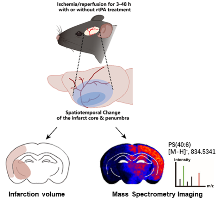
Mice were subjected to 60-minutes transient middle cerebral artery occlusion (tMCAO) with or without rtPA treatment and the brain tissues were collected for TTC staining or DESI-MSI at 3, 6, 9, 12, 24 and 48 hours reperfusion after tMCAO. (Image by Prof. CHANG Junlei)
Media Contact:
ZHANG Xiaomin
Email:xm.zhang@siat.ac.cn