Enhancing PET Image Quality with Deep Learning: A Cost-Effective Approach
Date:20-09-2023 | 【Print】 【close】
In the realm of positron emission tomography (PET), the axial field of view (AFOV) plays a pivotal role in determining image quality. While total-body PET scanners like the uEXPLORER offer superior sensitivity, they come at a higher cost and limited accessibility.
However, a novel method, spearheaded by Prof. HU Zhanli and a research team from the Shenzhen Institute of Advanced Technology (SIAT) at the Chinese Academy of Sciences, offers a solution to elevate PET image quality. Their approach leverages deep learning technology to optimize short-axis PET scanner images using high-quality long-axis PET scanner images.
The result was published in European Journal of Nuclear Medicine and Molecular Imaging on Sep. 06.
Their study involved conducting experiments on PET images from three anatomical locations (brain, lung, and abdomen) taken from 335 patients. Employing a well-established 3D neural network, the researchers generated short-axis images of comparable quality to long-axis images.
The results were impressive, with the proposed approach achieving superior image quality metrics in all three anatomical locations (peak signal-to-noise ratio exceeded 35 dB, 33 dB, and 38 dB, respectively, with statistical metrics P-values of less than 0.05). Moreover, both subjective physician evaluations and quantitative numerical analyses demonstrated the potential of this method to enhance short-axis PET image quality.
This study sheds light on the possibility of using the uEXPLORER PET/CT system to improve short-axis PET image quality economically. Additionally, it highlights the potential benefits for patients and radiologists through computer-aided diagnosis systems.
"Our study presents a cost-effective solution for hardware upgrades, streamlines the dissemination and implementation of software algorithm models, and paves the way for novel perspectives on the advancement and evolution of interconnected biomedical instruments," said Prof. HU.
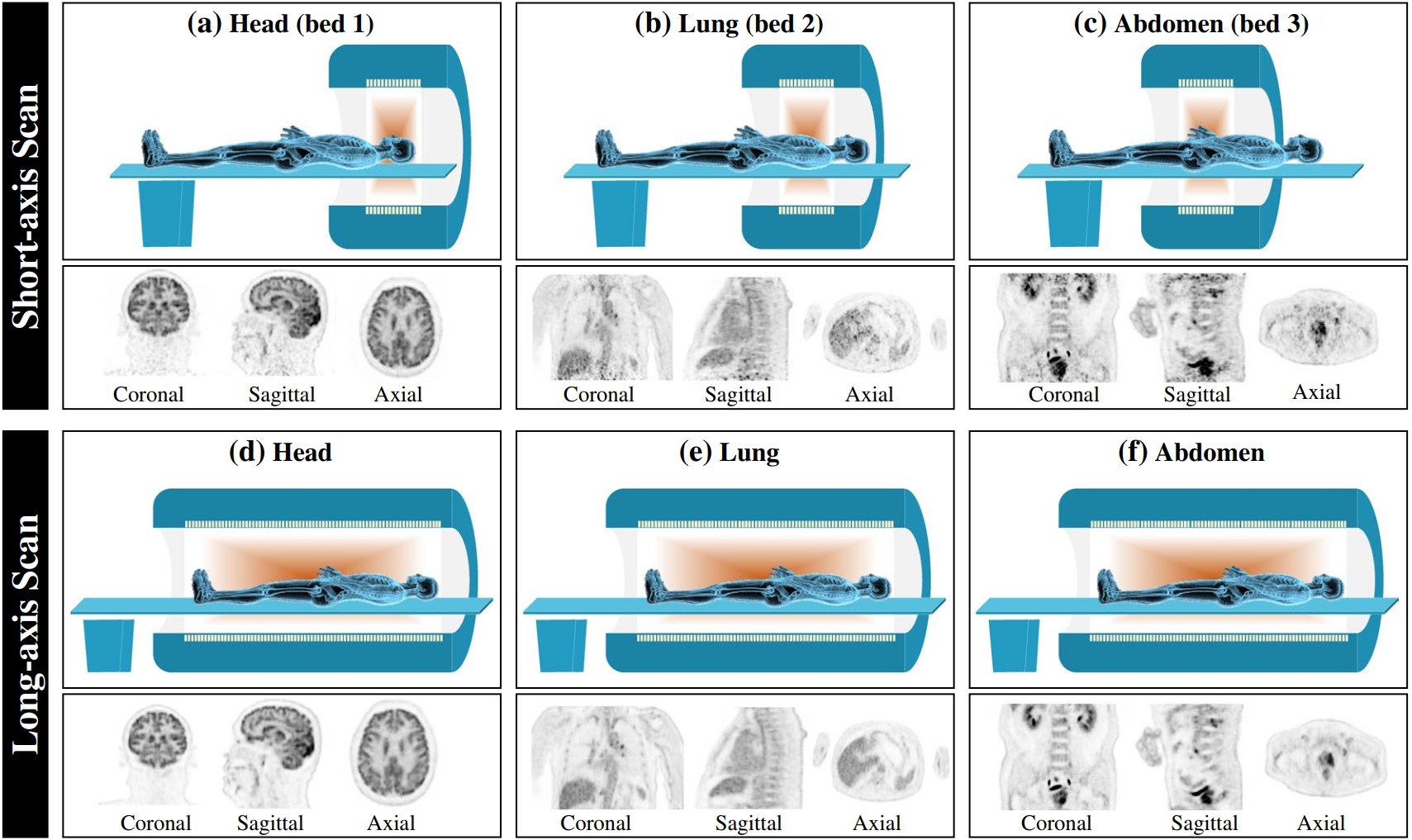
Illustration of long-axis images and short-axis images for 3 beds, including head, lung and abdomen. (Image by SIAT)
Media Contact:
ZHANG Xiaomin
Email:xm.zhang@siat.ac.cn