Head-Mounted Microscope Reveals High-Resolution Neurovascular Dynamics in Freely Behaving Mice
Apr 14, 2025
A research team led by Prof. LIU Chengbo from the Shenzhen Institutes of Advanced Technology (SIAT) of the Chinese Academy of Sciences, has developed a groundbreaking 1.7-gram head-mounted microscope. This innovative device can simultaneously capture neural activity and cerebral hemodynamics in freely moving mice, providing an unprecedented tool for exploring neurovascular coupling (NVC) in the brain and studying neurological disorders.
The study was published in Science Advances on March 21.
NVC represents the close temporal and regional relationship between neural activity and cerebral blood flow and oxygenation. When neural activity is initiated in a specific brain region, the metabolic demand within that region increases, leading to enhanced blood flow to supply greater amounts of oxygen and glucose, thus meeting the elevated metabolic needs of neurons. Conversely, if pathological changes hinder the cerebral vessels' capacity to provide sufficient oxygen and energy, the functional activity of the corresponding brain region will be significantly affected.
Conventional NVC imaging techniques face challenge to achieve simultaneous in vivo high spatiotemporal resolution of neuronal activity and cerebral blood flow. This limitation makes it difficult to accurately capture the dynamic relationship between neural activity and local hemodynamic changes. Additionally, most existing studies use head-fixed setups, which do not reflect true neurovascular coupling during natural animal behaviors.
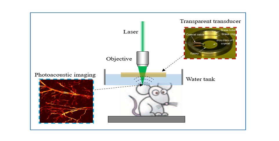
In this study, the researchers integrated confocal fluorescence microscopy (CFM) and photoacoustic microscopy (PAM) into a single miniaturized imaging probe, weighing only 1.7 grams. This dual-modal imaging approach achieves simultaneous imaging of neuronal burst firing and multi-parametric hemodynamics such as vascular oxygen saturation, at 0.78 Hz, with 1.5 μm lateral resolution over a 400 μm × 400 μm field of view.
To validate the NVC imaging capabilities of the microscope, the researchers conducted experiments on freely moving mice across various physiological states, including prolonged systemic hypoxia, localized rapid sensory stimulus, and acute epilepsy conditions. In these tests, they identified cell type-specific excitatory and inhibitory neurovascular coupling response to hypoxic challenges, observed the active role of arterioles in maintaining a safe oxygenation level in the somatosensory cortex during sensory stimulation, and detected abnormal oxygen depletion and vasodilatation preceding burst neuronal discharges in epileptic episodes.
"Our microscope, for the first time, enables dual-modal imaging at high spatiotemporal resolution, enhancing our understanding of neurovascular coupling mechanisms without affecting the natural behavior of the mice," said Prof. LIU.
This work holds significant potential for revealing the pathomechanisms of various brain diseases.

Head-mounted dual-modal microscope and mounted on a mouse brain (Image by CHEN Ningbo)
File Download:
Media Contact
LU Qun
Email:
qun.lu@siat.ac.cn
qun.lu@siat.ac.cn
More on this News Release
JOURNAL
Science Advances
DOI
10.1126/sciadv.adu1153
Related Articles
Dec 17, 2024
Researchers develop an ultrasensitive, broadband transparent ultrasonic transducer based on a novel transparent piezoelectric single crystal, achieving high-resolution, large-field-of-view, and rap...