New Cell-scale Method Reconstructs Whole-brain Fiber Tracts from Routine Histology
Nov 04, 2025
Recently, a research team led by Prof. XU Fang, Prof. LIU Cirong Liu, Prof. XIAO Yanyang, and Prof. BI Guoqiang from the Shenzhen Institutes of Advanced Technology (SIAT) and the Center for Excellence in Brain Science and Intelligence Technology (CEBSIT) of the Chinese Academy of Sciences (CAS) reported a novel method that infers axonal pathways from cytoarchitectonic orientation to reconstruct three-dimensional whole-brain fiber tracts at cellular scale in primate and human tissues. The study, entitled "Whole-brain reconstruction of fiber tracts based on cytoarchitectonic organization," was published in Nature Methods on November 3, 2025.
The method, termed CABLE (cytoarchitecture-based link estimation), recovers complex crossings and sharp turns from routine Nissl or DAPI stains and agrees with gold-standard viral tracing.
Mapping how fiber bundles connect brain regions is essential for understanding function and disease. Existing tools force a trade-off between coverage and detail. Diffusion MRI is high-throughput but coarse. Viral tracing reaches micrometers but lacks whole-brain scalability. CABLE is designed to bridge this gap.
Although routine histology visualizes cell bodies rather than axons, local cell shapes show non-random orientation that aligns with nearby fiber bundles. CABLE leverages this anisotropy to infer tract directions from standard stains.
Built on the VISoR high-throughput volumetric imaging platform, the workflow sections whole brains at 300 μm, clears and stains tissue with DAPI or Nissl, acquires volumetric data, stitches it, then computes orientation distribution functions for 3D tractography without genetic labels or viruses. The method is cross-species and platform-agnostic.
CABLE reconstructs densely packed, intersecting, and sharply bending fibers in macaque and marmoset brains and validates trajectories against viral-tracer datasets. In neonatal hypoxic-ischemic encephalopathy samples, it detects abnormal corpus callosum fibers, suggesting diagnostic value for neuropathology.
Overall, CABLE outperforms ex vivo diffusion MRI in resolving complex geometries while avoiding the low throughput of viral tracing. It offers a practical route to high-precision, high-throughput connectome mapping with potential applications in psychiatry and epilepsy.
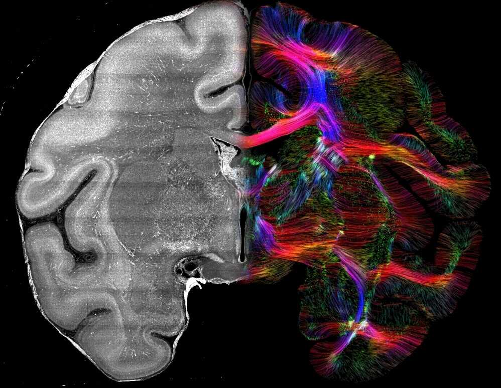
Nissl staining and CABLE-reconstructed fibers. The left panel shows a Nissl-stained section of a macaque brain, while the right panel displays the fiber map reconstructed from the same Nissl-stained image using CABLE. Color codes indicate different directions: red for left–right, green for anterior–posterior, and blue for dorsal–ventral orientation. (Image by SIAT)
File Download: